Facilities and Lab Equipment
Our lab is currently under construction. Our current lab systems include thin film fabrication and characterization facilities. Koç University Surface Technologies Research Center (KUYTAM) provides the essential materials characterization equipment in the shared experimental facilities. Koç University is also currently constructing new central shared experimental cleanroom and characterization facilities, which is going to contain standard optical and electron beam lithography tools, maskless lithography, deposition systems, etching instruments and the rest of the necessary fabrication and characterization equipment for electronic and magnetic devices. The cleanroom is expected to be operational in 2018.
In addition to the shared facilities, we are currently setting up our own thin film growth and characterization labs, as described below:
Thin film growth instruments
- Pulsed Laser Deposition
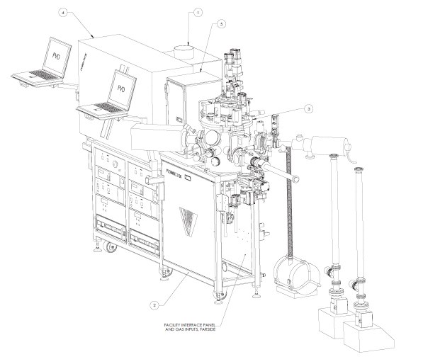
Figure 1. Pulsed laser deposition setup (Photo credit: PVD tools)
Pulsed laser deposition setup is going to be operation in 2018. This instrument is going to be used for growing functional oxide thin films with good thickness and stoichiometry control. These functional oxides are going to be used for experiments on defect chemistry-mediated magnetism, voltage-controlled magnetism and other hysteresis behavior, piezoelectricity, ferroelectricity and magneto-optical Faraday and Kerr rotation materials. Instrument details are going to be announced later.
- Sputtering system
Sputtering system is useful particularly for depositing magnetic oxides, magnetic or other functional metals as well as contacts for electrical transport measurements. Instrument details are going to be announced later.
Thin film characterization instruments
- Room temperature Faraday and Kerr rotation measurement setup
Currently, we are constructing Kerr rotation setup for room temperature measurements of magneto-optical Kerr rotation as a function of magnetic field at room temperature.
- Brillouin light scattering microscope
We are constructing frequency, phase and spatially-resolved Brillouin light scattering microscope for imaging spin waves between 2-20 GHz, which are excited using grounded coplanar waveguides.
- Multiferroic tester (Radiant, P-H, P-V loop tester)
- Vibrating sample magnetometer (ready in Fall 2018)
- Magnetoelectric transport measurement setup and probe station (ready in Fall 2018)
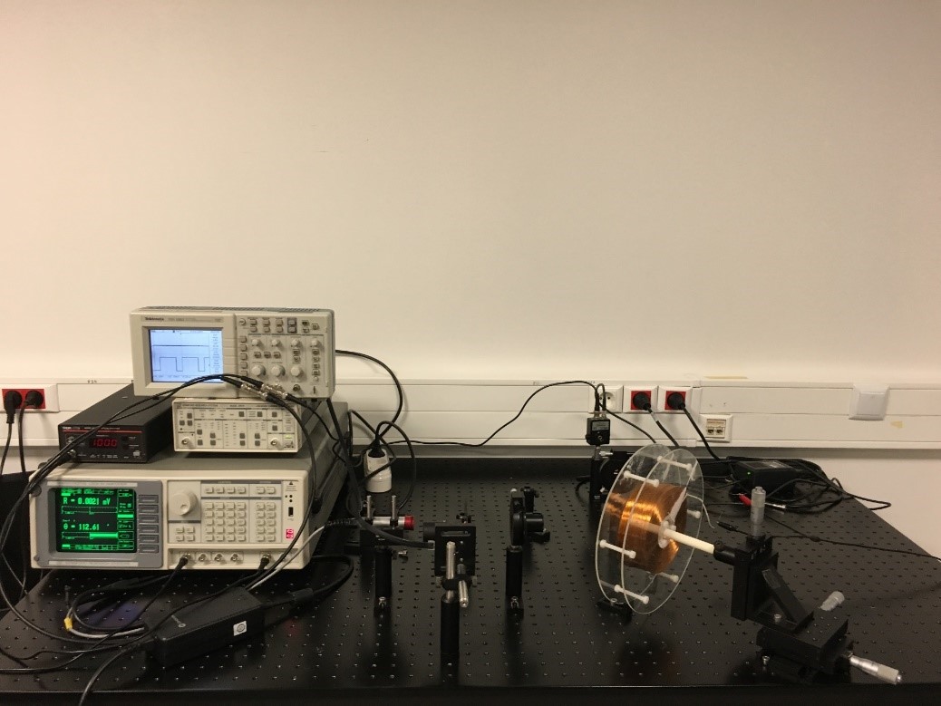
Figure 2. Kerr rotation setup (Photo credit: Soheila Kharratiankhameneh)
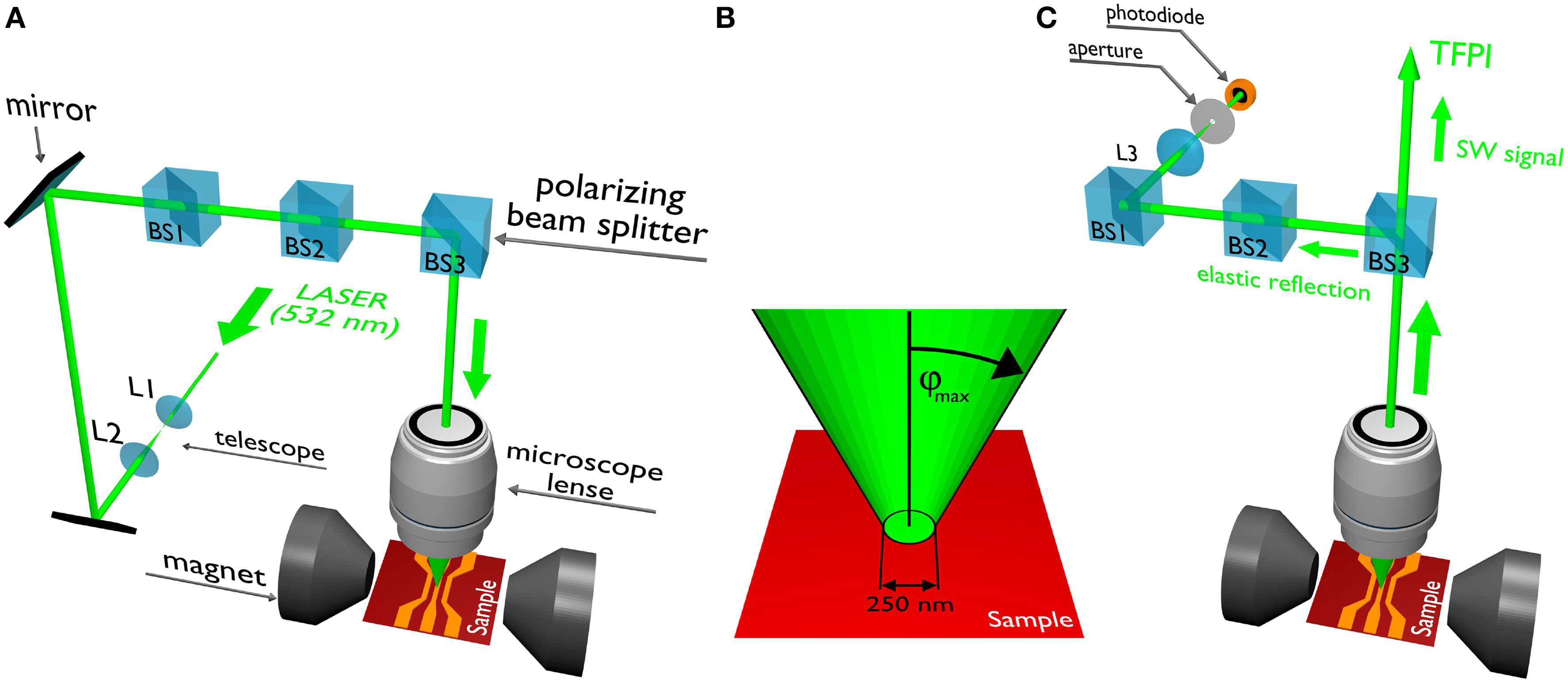
Figure 3. Schematic drawing of the Brillouin light scattering setup which we are currently building [1]
References
- Sebastian, K. Schultheiss, B. Obry, B. Hillebrands and H. Schultheiss, “Micro-focused Brillouin light scattering: imaging spin waves at the nanoscale,” Frontiers in Physics 3:35 (2015). doi: 10.3389/fphy.2015.00035